Brain Is Two Separate Organs: Stanford Study Overturns Decades of Neuroscience
Two Separate Organs: The Brain's Hidden Duality
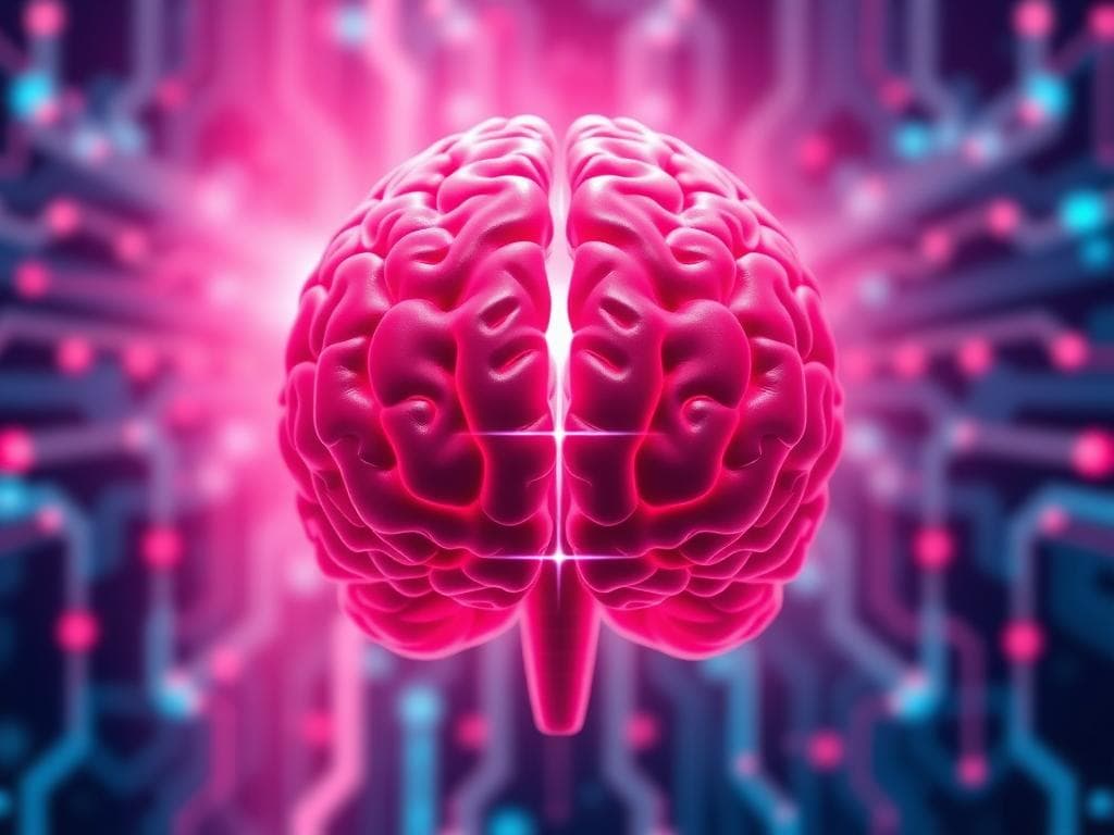
For centuries, neuroscience has treated the brain as a single, unified organ. But a landmark study from Stanford Medicine, published September 18 in Nature Neuroscience, reveals a far more complex reality: the human brain is actually two distinct organs that evolved independently over hundreds of millions of years. The discovery challenges a foundational model of brain development and explains decades of frustrating failures in laboratory research.
Led by associate professor of developmental biology Kyle Loh, the research team identified two completely separate progenitor cell populations during embryonic development—one destined to become the forebrain and midbrain, the other committed to forming the hindbrain. These populations never overlap, running on parallel developmental tracks that remain mutually exclusive from the earliest stages of life.
A Fundamental Split Found in Embryonic Development
The breakthrough came from studying gastrulation, the earliest stage when the body first takes shape. Graduate students Carolyn Dundes and Rayyan Jokhai examined developing mouse embryos and discovered that the hindbrain follows a developmental path entirely separate from the forebrain and midbrain—not branching off from a common ancestor as previously believed.
The researchers identified two distinct progenitor cells: one expressing the Otx2 gene, destined for forebrain and midbrain, and another expressing Gbx2, committed to hindbrain formation. These two populations remain completely separate from the earliest stages. Examination of chromatin—the DNA packaging that determines gene accessibility—revealed fundamentally different configurations in each progenitor, effectively locking each into its respective fate.
Explaining Decades of Failed Lab Experiments
This discovery resolves a long-standing mystery in stem cell biology. For decades, scientists have struggled to grow human hindbrain neurons in the laboratory, despite successfully cultivating other brain cell types. The reason, as Jokhai explains, is that "previous attempts to make hindbrain neurons likely tried to coax forebrain and midbrain progenitors into hindbrain cells, which our study shows is not possible."
Armed with this new understanding, the Stanford team successfully coaxed human pluripotent stem cells to become functional hindbrain motor neurons for the first time. These lab-grown neurons exhibited authentic characteristics, including action potentials and proteins identifying the hindbrain segments that control facial and swallowing muscles, validating the new developmental model.
Evolutionary Origins Spanning 550 Million Years
The two-brain pattern extends far beyond humans. The researchers found the same dual-origin structure in chickens, zebrafish, and even acorn worms—tiny ocean-floor creatures sharing a distant ancestor with humans. Jellyfish, which diverged from humans 600 to 700 million years ago, possess two nervous systems at opposite ends of their bodies, suggesting this dual structure is deeply ancient.
"Our research suggests that evolution took two existing neural systems and pushed them together spatially," Loh said. "Having the brain as one organ would probably be more efficient, but we rely on this primordial way to make the brain as two separate pieces."
Implications for ALS, SMA, and Brain Stem Diseases
The discovery carries profound clinical implications. The hindbrain controls essential functions like breathing, heartbeat regulation, swallowing, and hunger. Diseases like spinal muscular atrophy (SMA)—a leading genetic cause of death in children under 1—and amyotrophic lateral sclerosis (ALS) specifically affect hindbrain neurons, causing patients to lose the ability to swallow and breathe.
Until now, studying these diseases has been nearly impossible because scientists cannot obtain brain stem tissue from living patients. The ability to grow authentic hindbrain neurons in a dish opens unprecedented opportunities for understanding what goes wrong in these conditions and developing regenerative therapies.
There's also an unexpected connection to obesity treatment: the hindbrain contains circuits regulating hunger, which is precisely how weight-loss drugs like semaglutide work. This research could accelerate development of more targeted treatments for metabolic disorders as well.
Beyond the Single-Organ Paradigm
The study overturns decades of scientific consensus rooted in the single-progenitor model. "We've shown for the first time that the front of the brain arises from a totally different progenitor cell than the back of the brain," Loh said. "Our discovery means that we can now grow neurons from the back of the brain, the hindbrain, in a petri dish and study their functions."
The researchers plan to extend their work to determine the developmental origins of the spinal cord and investigate exactly how SMA and ALS compromise hindbrain neuron function. This research, supported by the National Institutes of Health, the California Institute for Regenerative Medicine, and the Spinal Muscular Atrophy Foundation, among others, represents a fundamental shift in understanding our most complex organ.
As Jokhai noted, "Even 500 million years ago, there were these separate neural systems, which now almost operate as one, which is very cool. Now we have a model to better understand these devastating diseases, and work toward regenerative therapies for them."
Related News

LLM Writing Guide: Rules, Tools, and AI's Expanding Role

Claude-Assisted Exploit Chain Compromises OpenAI Internal Repos

Bend Language: Prove Code Correct, Run Fast on GPU

4B Model Beats Postgres Query Optimizer by 81%
TypeSafe's Jev: A New AI Model for Ultra-Fast, Zero-Hallucination Decisions